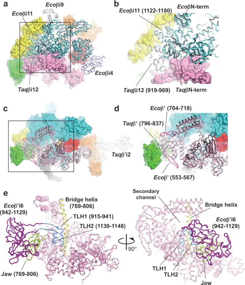
FIGURE 2.
Structure comparisons of the β and β′ subunits of the E. coli and T. aquaticus RNAPs. a, superposition of Ecoβ and Taqβ RNAPs. Ecoβ (cyan) and Taqβ (black) are shown as α-carbon backbones in addition to the molecular surfaces of other E. coli RNAP subunits (αI, yellow; αII, green; β′, pink; σ70, orange; and σ1.1, red). b, magnified view of the boxed region in a. c, superposition of Ecoβ and Taqβ RNAPs. Ecoβ′ (pink and magenta), Taqβ′ (black and white) are shown as α-carbon backbones in addition to the molecular surfaces of other E. coli RNAP subunits (αI, yellow; αII, green; β′, pink; σ70, orange; and σ1.1, red). d, magnified view of the boxed region in c. e, the bridge helix (yellow), TLH (light blue), and jaw (yellow green) are highlighted on the α-carbon backbone of the Ecoβ′ (pink) structure. Ecoβ′i6 (purple) was modeled using the E. coli core enzyme model (24).

