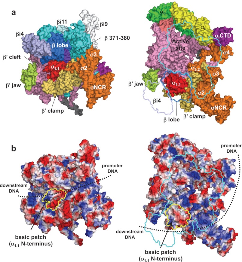
FIGURE 4.
Structure and function of σ1.1. a, molecular surface of the holoenzyme with σ1.1. Left, front view; right, side view. In the right panel, β subunit has been removed and outlined for clarity. b, electrostatic distribution of the holoenzyme. Left, front view; right, side view (orientations are the same as in a). Positive electrostatic potential is blue, and negative potential is red. The positions of σ1.1 in these views are indicated by yellow outlines. A basic patch found at the σ1.1 N terminus is shown. The potential DNA pathway during open complex formation is shown by dotted lines.

