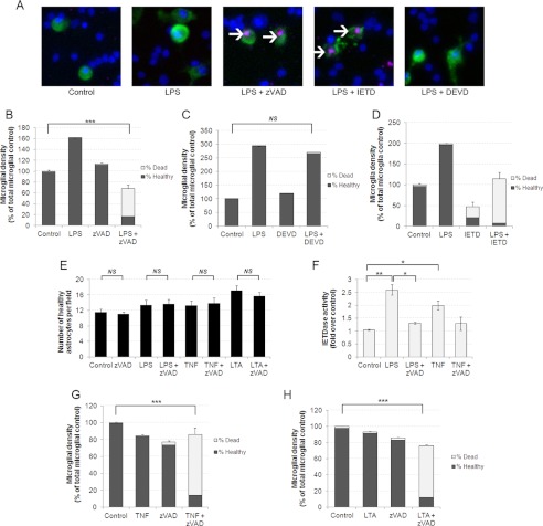
FIGURE 1.
Inhibition of caspase-8 selectively kills microglia following inflammatory activation. A, caspase inhibitors Z-VAD and IETD induce necrosis of microglia in mixed neuronal glial cultures. Microglia are labeled in green using Alexa Fluor 488-labeled isolectin-B4, nuclei are labeled in blue using Hoechst 33342, and necrotic cells have pink nuclei due to PI labeling. Necrotic microglia are indicated with white arrows. B–D, Z-VAD (50 μm) and IETD (50 μm) but not DEVD (50 μm) induce death of microglia in the presence of LPS. Microglia were treated for 48 h with 100 ng/ml LPS and then for a further 24 h with indicated caspase inhibitor before quantifying death. Data are means ± S.E. of % dead microglia (n = 3). E, astrocytes are not killed by caspase-8 inhibitors and inflammatory activation. Astrocytes were quantified by nuclear morphology following the same treatments as described in B–D. LTA was used at 50 μg/ml, TNF-α was added at 50 ng/ml. Data are means ± S.E. (n = 3). F, Z-VAD inhibits LPS- and TNF-α-induced IETDase cleavage activity in pure microglia. Pure microglia were treated with LPS or TNF-α for 24 h followed by 2 h in the presence of Z-VAD (50 μm). IETDase activity was then measured in cell extracts. Data represent mean ± S.E. (n = 4). G and H, TNF-α and LTA treatment render microglia susceptible to Z-VAD-induced necrotic death (n = 3). NS, not significant. * = p < 0.05, ** = p < 0.01, *** = p < 0.001.

