
FIGURE 5.
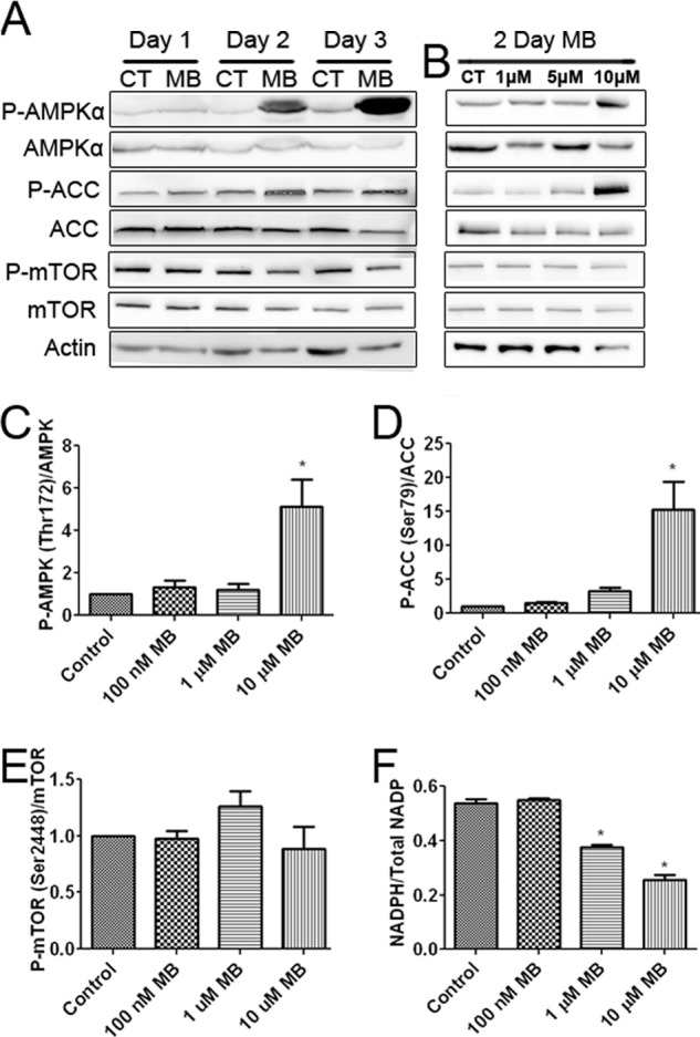
MB activates AMPKα and inhibits ACC. A, Western blots are shown of ACC, AMPKα, and mTOR after 1, 2, and 3 days of 10 μm MB treatment compared with control. MB increased the phosphorylation of AMPKα and ACC while mildly decreasing that of mTOR. CT, control. B, Western blots of AMPKα, ACC, and mTOR after 2 days of MB treatment at the indicated concentrations are shown. MB, at 10 μm, increased AMPKα and ACC phosphorylation and mildly decreased mTOR phosphorylation. C, densitometry of P-AMPK/AMPK ratio after 2 days of MB treatment is shown. 10 μm MB significantly increased the ratio of P-AMPKα/AMPK. D, densitometry of p-ACC/ACC ratio after 2 days of MB treatment is shown. 10 μm MB significantly increased the ratio of p-ACC/ACC. E, densitometry of P-mTOR/mTOR ratio after 2 days of MB treatment is shown. MB had no effect on the ratio of P-mTOR/mTOR. F, shown is the cellular NADPH to total NADP ratio. Concentrations of 1 and 10 μm MB significantly decreased NADPH/Total NADP compared with control. * indicated p < 0.05 by 1-way ANOVA and Tukey's post-hoc test.