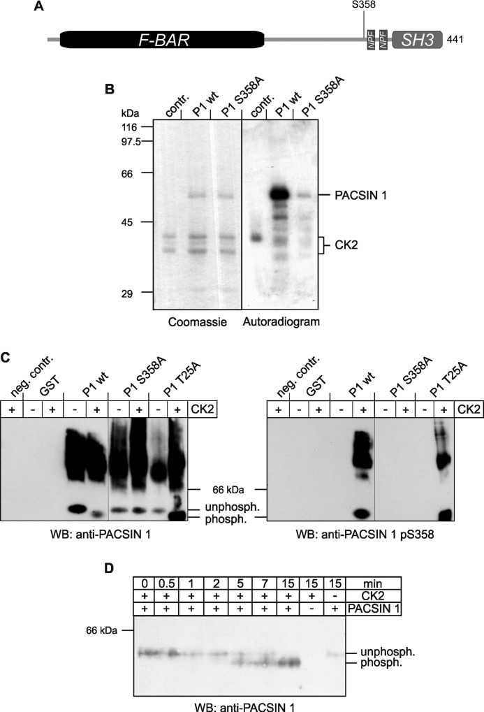
FIGURE 1.
Phosphorylation of PACSIN 1 at serine 358 by CK2. A, scheme of PACSIN 1 represents its domain structure and location of Ser358. B, recombinant wild-type PACSIN 1 (P1 wt) and PACSIN 1 S358A (P1 S358A) were purified as GST fusion proteins, and GST was removed by proteolytic cleavage. The proteins were incubated with CK2 and [γ-32P]ATP for 20 min and analyzed by SDS-PAGE. The SDS-polyacrylamide gel was stained with Coomassie Brilliant Blue, dried, and exposed to an x-ray film. C, recombinant proteins including GST and an unrelated mutant PACSIN 1 T25A (P1 T25A) as controls were incubated with (+) or without (−) CK2, resolved by native PAGE, and immunoblotted with antibodies specific for PACSIN 1 or PACSIN 1 pS358. D, for kinetic analysis wild-type PACSIN 1 (P1 wt) was incubated with CK2 for the indicated time periods, separated by native PAGE and immunoblotted (WB) for PACSIN 1.

