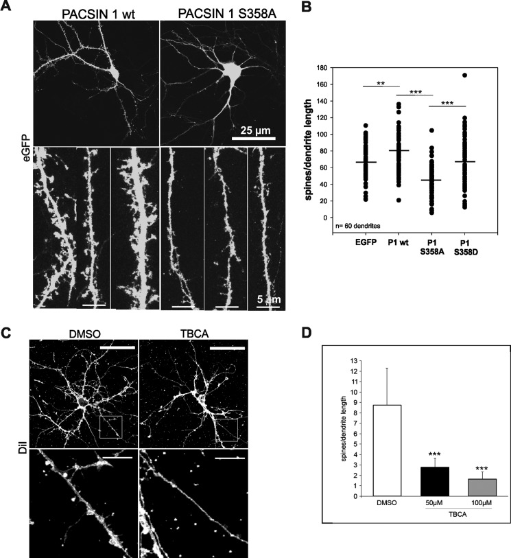
FIGURE 4.
Reduced spine formation in the presence of the PACSIN 1 S358A mutant or after CK2 inhibition. A, rat hippocampal neurons were transfected with either PACSIN 1 wt (left). the mutant protein S358A (right) or the mutant protein S358D (data not shown) in combination with EGFP to visualize spines (upper panel). Enlargements of three dendrite segments of neurons transfected with either PACSIN 1 wt (left) or S358A mutant (right) are shown. Fewer spines can be detected in neurons expressing PACSIN 1 S358A (lower panel). B, quantification of two independent experiments. For each group five dendrites were counted on each of 12 cells, n = 60 dendrites; **, p < 0.01; ***, p < 0.001, t test. C, untransfected hippocampal neurons were treated either with dimethyl sulfoxide (DMSO) or the CK2 inhibitor TBCA in dimethyl sulfoxide and stained with the DiI plasma membrane dye to visualize the spines (upper panel). Enlargements of the indicated dendrite segments of neurons are shown (lower panel). D, quantification of three independent experiments is shown. For each group three dendrites were counted on each of five cells, n = 45 dendrites; ***, p < 0.001, t test. Error bars, S.D.

