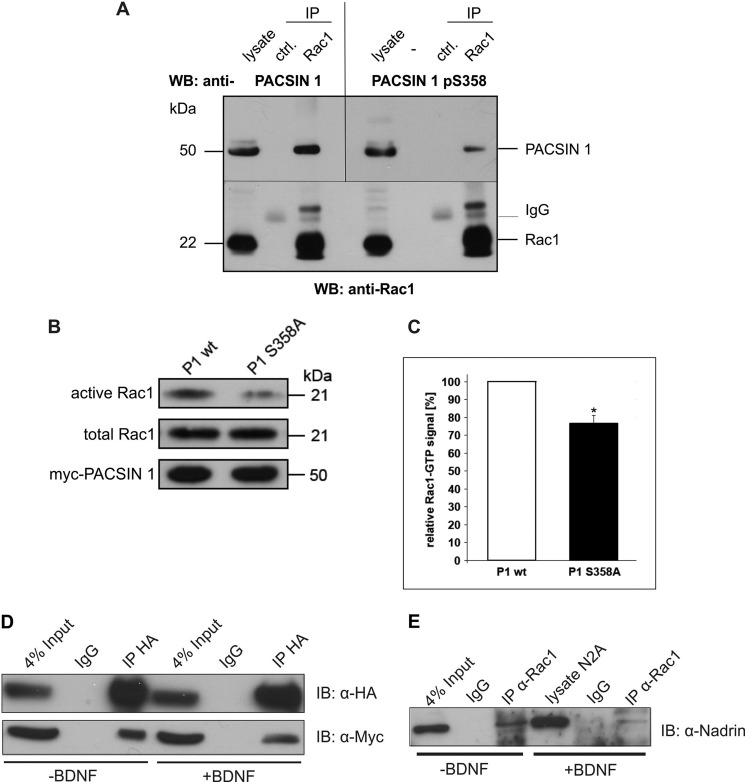
FIGURE 5.
PACSIN 1 pS358 is present in a complex with the spine regulator Rac1 and the GAP NADRIN. A, mouse brain homogenate was used for immunoprecipitation (IP) with an antibody specific for Rac1 (lower panel) and co-precipitated proteins analyzed by immunoblotting (WB). The bands marked IgG represent immunoglobulin light chains. Total PACSIN 1 (upper panel left) as well as PACSIN 1 pS358 (upper panel right) are detected in the Rac1-IP lane. B, the levels of GTP-bound Rac1 were determined in PACSIN 1 wt or PACSIN 1 S358A-transfected N2a cells which were lysed 24 h after transfection. Pulldown experiments were performed, and equal amounts of bead fractions and lysate fractions were resolved on SDS-polyacrylamide gels (15%). The GTP-bound forms of Rac1 in bead fractions and total Rac1 in lysate fractions were stained with mouse anti-Rac1 antibody (top and middle panels). Transfection efficiencies of both constructs were comparable (bottom panel). C, quantification of three independent experiments was performed using ImageJ. *, p < 0.05, t test. Error bar, S.D. D, N2a cells were co-transfected with hemagglutinin (HA)-tagged NADRIN and Myc-tagged PACSIN 1 wt, treated with (+) or without (−) BDNF (100 ng/ml) for 5 h, and lysed. NADRIN was precipitated with an antibody specific for HA, and comparable amounts of the individual precipitates were resolved by SDS-PAGE. After immunoblotting (IB), the precipitated proteins were visualized with antibodies specific for the indicated tags. E, N2a cells were treated with (+) or without (−) BDNF (100 ng/ml) for 5 h and lysed. Endogenous Rac1 was precipitated with a specific antibody, and comparable amounts of the individual precipitates were resolved by SDS-PAGE. After immunoblotting co-precipitated endogenous NADRIN was visualized with a specific antibody.

