FIGURE 1.
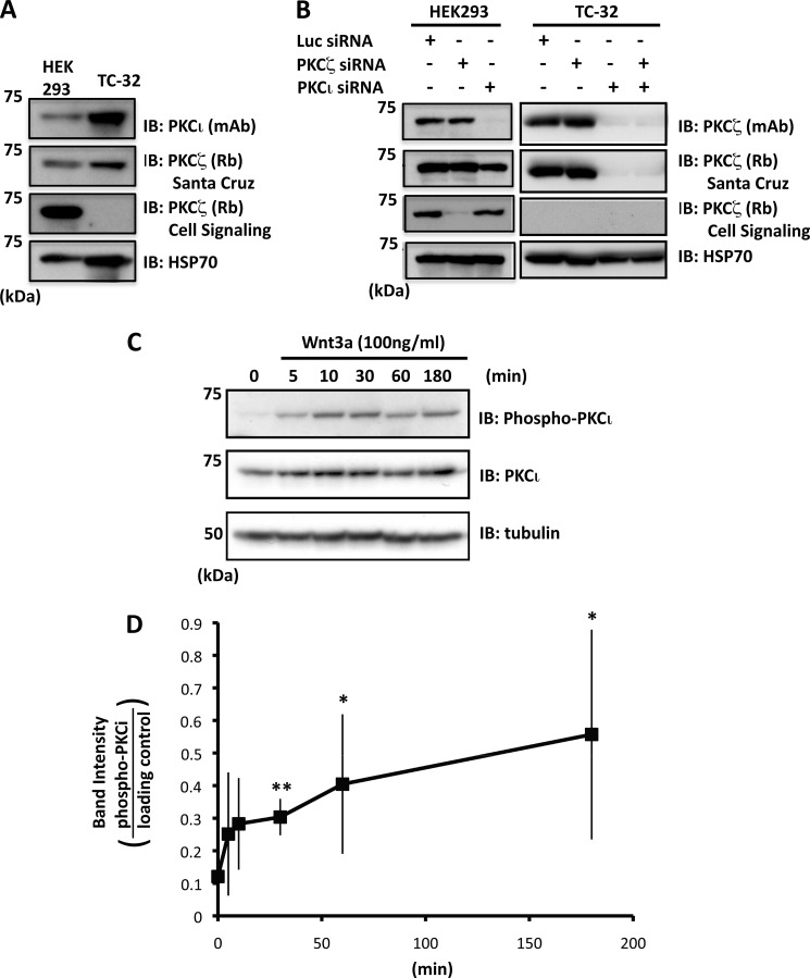
PKCι is expressed by TC-32 cells and phosphorylated in response to Wnt3a. A, immunoblot analysis of PKCι and PKCζ in HEK293 and TC-32 cell lysates. PKCι was detected in TC-32 cells, but contradictory results were obtained for PKCζ with two different antibodies. B, combination of immunoblot analysis and isoform-specific siRNA indicated that PKCι, but not PKCζ, is expressed by TC-32 cells. PKCζ antiserum from Santa Cruz reacts with both PKCζ and PKCι. Note that knockdown in HEK293 confirmed the specificity of the PKCι and PKCζ siRNA reagents. C, time course of PKCι phosphorylation stimulated by Wnt3a (antibody recognizes Thr412 in PKCι and Thr410 in PKCζ). D, quantitative results of Wnt3a-dependent PKCι phosphorylation. The band intensity of phospho-PKCι was normalized to loading control (HSP70 or tubulin). The graph shows average values ± S.D. of five independent experiments. *, p < 0.05; **, p < 0.01 compared with zero time point. Similar results were obtained when data were normalized to total PKCι. However, because of fluctuation in total PKCι levels, the statistical significance was evident only at the 3-h time point. IB, immunoblot.

