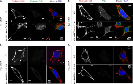
FIGURE 2.

Immunofluorescent analysis of pPKCι and total PKCι in TC-32 cells with or without Wnt3a treatment. The cells were transfected with luciferase (Luc; A and C) or PKCι (B and D) siRNA reagents and 48 h later incubated with serum-free culture medium ± Wnt3a (100 ng/ml) for 1 h, fixed in formaldehyde, and then stained for phalloidin, DNA (DAPI), and pPKCι (A and B) or total PKCι (C and D). A scale bar is indicated. Ctl., control.
