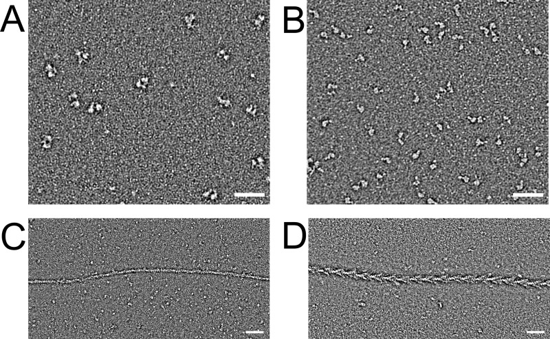
FIGURE 3.
Electron microscopic images of myosin-18A fragments. A, field of negatively stained myosin-18Aα HMM molecules. Scale bar, 50 nm. B, field of negatively stained myosin-18Aα-S1 molecules. Scale bar, 50 nm. C, myosin-18Aα-S1 mixed with equimolar actin in the absence of nucleotide demonstrates the weakness of actin binding. D, nonmuscle myosin-2A-S1 mixed with actin under the same conditions shows the classic arrowhead decoration.

