
Fig. 2.
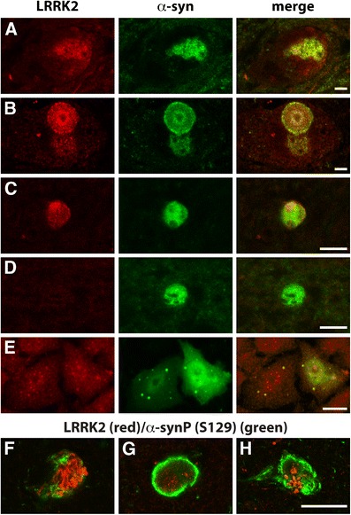
Co-localization of LRRK2 and α-synuclein in PD brain and cell models. In PD brains (a–d, f–g), merged images clearly outline single neurons in the substantia nigra (a, b) and Lewy bodies (b–d, f–g) using double-labelling immunofluorescence. There is an increase of LRRK2 and α-synuclein immunoreactivity in brainstem neurons without Lewy body formation (a), with LRRK2 co-localizing with α-synuclein in Lewy bodies (donut inclusion in b) in these neurons. The co-localisation of LRRK2 and α-synuclein was also observed in cortical Lewy bodies (c). Cortical Lewy bodies without LRRK2 immunoreactivity were also observed (d). S129 phosphorylated α-synuclein antibody also confirmed co-localisation of LRRK2 with phosphorylated α-synuclein, with LRRK2 often centralized to a radiating pattern of phosphorylated α-synuclein fibrils (f–h). In the H4 cell model, double-labelling immunofluorescence for α-synuclein inclusion formation shows that endogenous LRRK2 co-localizes with α-synuclein inclusions (e). Scales in all panels are equivalent to 10 μm