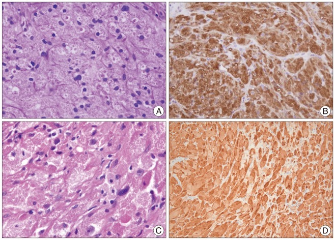
Fig. 2.
Histologic examination. A and C : Cases 1 (A) and 2 (C) show granular cells with abundant eosinophilic granules in the cytoplasm. Hyalinizing fibrosis is also observed in all cases. Some lymphocytes have infiltrated the tumor. There is mild nuclear pleomorphism but no necrosis. B and D : The tumor cells are strongly and diffusely immunoreactive with S-100 protein in Case 1 (B) and Case 2 (D).

