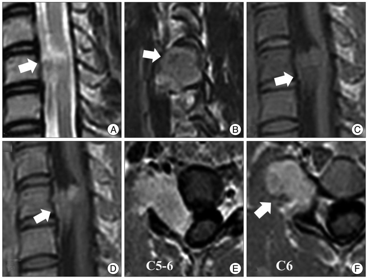
Fig. 3.
Preoperative magnetic resonance (MR) images. A : A T2-weighted MR image shows an isosignal intensity mass at C5-6. B : A T2-sagittal MR image shows a tumor extending to the right foramen. Speckled dots in the center of the tumor are of low signal intensity in a T2-weighted MR image (arrow). C and D : T1-weighted MR images show an isointense and well-enhanced mass. E and F : Axial MR images show that the spinal cord is displaced to the left by the tumor. The tumor is mainly located in the extraforaminal area. Low signal speckled dots are observed in all sequences of MR images.

