
Fig. 2.
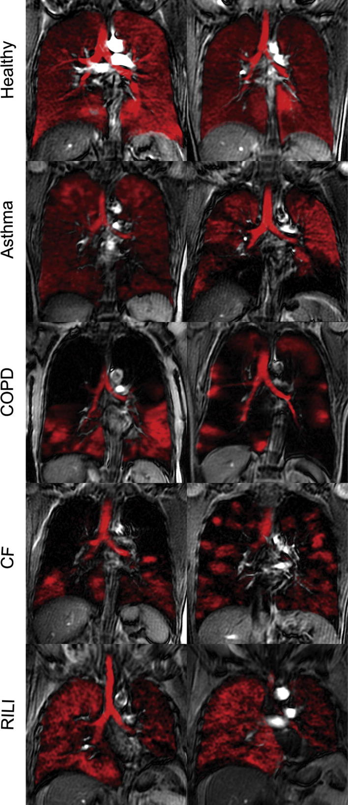
Hp 129Xe slice selective coronal MR images (in red) overlayed onto corresponding proton thoracic images from healthy volunteers and subjects with asthma, chronic obstructive pulmonary disease (COPD), cystic fibrosis (CF) and radiation-induced lung injury (RILI). Reprinted with permission from Shukla et al. Acad. Radiol., 2012; 19:944, © 2012 Elsevier. (For interpretation of the references to color in this figure legend, the reader is referred to the web version of this article.)