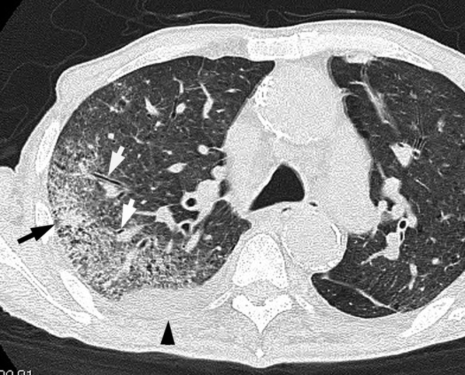
Figure 1.

Acute pneumonia caused by Pseudomonas aeruginosa in a 78-year-old male with a smoking habit and cardiac disease 3 days after the onset of fever, cough and dyspnoea. Transverse thin-section CT image (1-mm thickness) at the level of the tracheal carina shows bronchial wall thickening (white arrows) and consolidation (black arrow) in the right upper lobe. Right pleural effusion is present (arrowhead).
