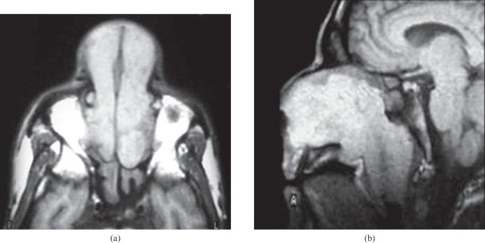
Figure 2.
Nasal scleroma. (a) Axial T1 weighted image shows a bilateral hyperintense nasal mass that extends through the anterior nares associated with retained secretions in the sphenoid sinus. (b) Sagittal T1 weighted image of another patient shows hyperintense signal intensity of the nasal mass that passes through the posterior choana and obliterates the nasopharyngeal airway.

