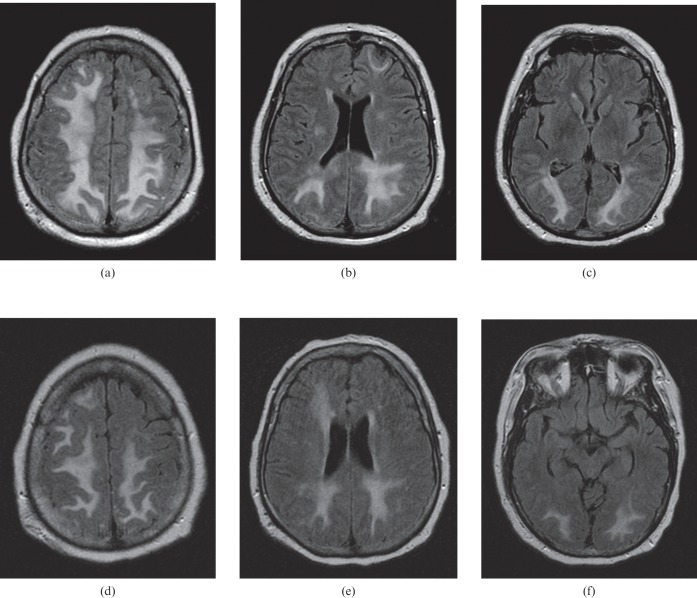
Figure 2.
A 61-year-old male presenting with tonic–clonic seizures, progressing to coma. (a–c) Brain MRI [fluid attenuated inversion recovery (FLAIR) sequence] obtained on the day of presentation demonstrates marked vasogenic oedema involving subcortical, deep and periventricular white matter of the frontal, parietal and occipital regions, while maintaining a predominant posterior pattern. (d–f) Brain imaging (FLAIR sequence) performed 6 days later demonstrates improvement of the vasogenic oedema.

