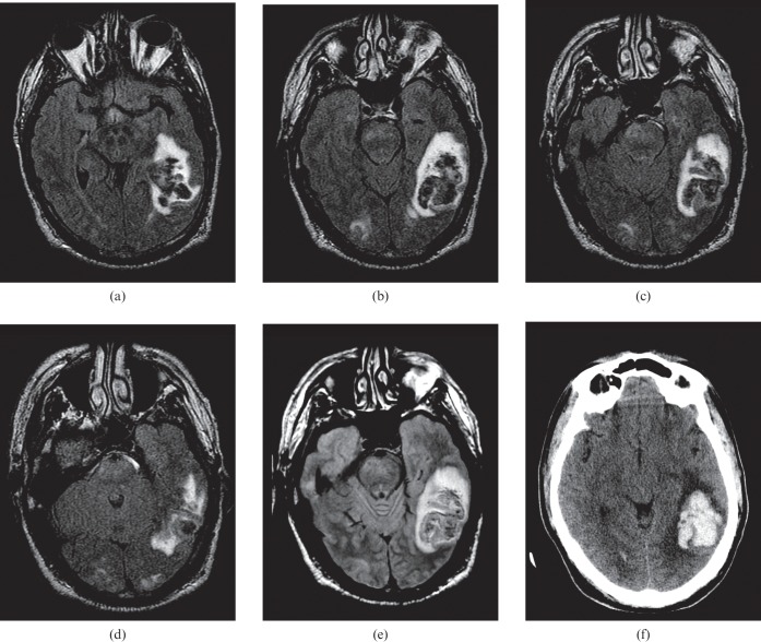
Figure 6.
A 39-year-old male with an appendiceal carcinoid tumour treated with chemotherapy complicated by haemolytic uraemic syndrome. Systolic blood pressure at the time of the study was >220 mmHg. (a–d) Brain MRI (fluid attenuated inversion recovery sequence) demonstrates numerous foci of vasogenic oedema in the occipital regions, cerebellum and brain stem. A large haematoma is seen in the left temporal lobe. (e) Proton density image demonstrates a haematoma in the left temporal lobe. (f) Non-contrast CT again demonstrates the known haematoma with surrounding vasogenic oedema.

