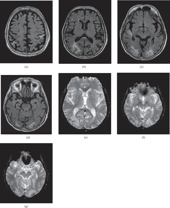
Figure 8.
An 87-year-old female presenting with a 15 min episode of visual changes and slurred speech. Blood pressure at the time of presentation was 176/99 mmHg. (a–d) Brain MRI (fluid attenuated inversion recovery sequence) demonstrates vasogenic oedema in the subcortical white matter of the left post-central gyrus, the right parietal lobe, right temporal lobe and bilateral occipital lobes. (e–g) T2* gradient sequence demonstrates haemosiderin in a petechial cortical gyral distribution.

