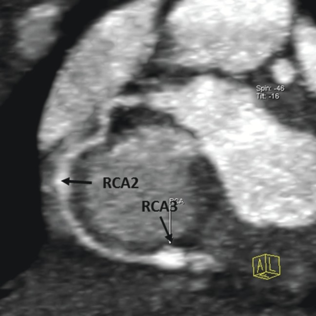
Figure 4.

Thin maximum intensity projection images showing the circumscribed dilations in the right coronary artery (RCA), middle segment (RCA2), and a circumscribed aneurysm in the RCA, distal segment (RCA3); the aneurysm in the RCA2 was not detected by transthoracic echocardiography.
