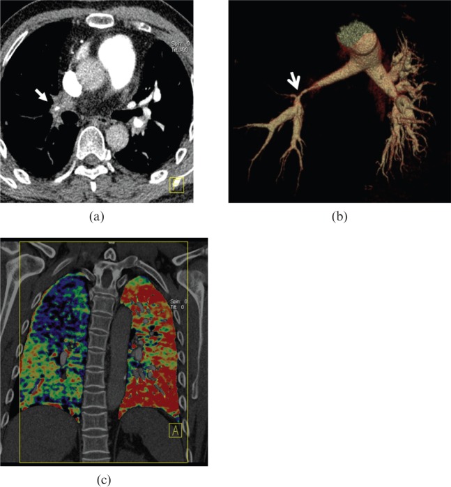
Figure 12.
A 41-year-old male with Takayasu arteritis. (a) Axial post-enhanced CT image shows the right lower lobe pulmonary artery wall is significantly thickened with a severely narrowed lumen [arrows in (a) and (b)]. (b) Volume-rendered image exhibits the significantly stenotic lesion in the right pulmonary artery trunk with decrease in blood perfusion of the right lung, compared with the left one in the dual energy CT iodine mapping image (c).

