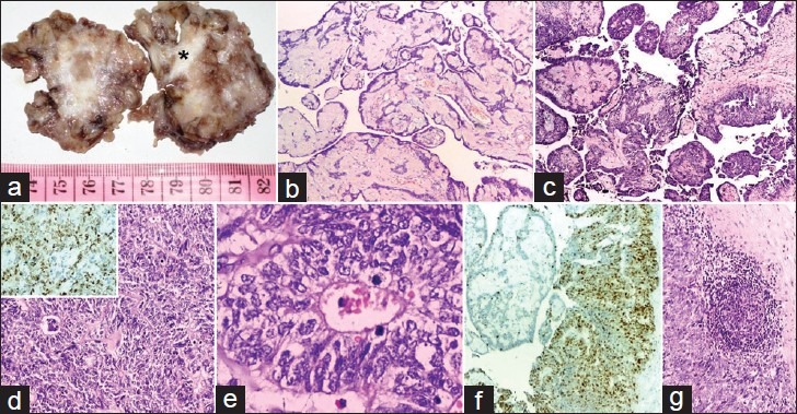
Figure 2.

(a) Cut-surface with solid-cystic areas and enclosed coccyx (*); (b) papillae with myxoid fibrovascular cores lined by benign ependymal cells (H and E, ×100); (c) myxopapillary component in continuity with anaplastic ependymoma component (H and E, ×100); (d) composed of perivascular rosettes and canals (H and E, ×100; inset: High Ki-67 labeling index); (e) lined by pleomorphic cells with nuclear atypia and mitotic figures (H and E, ×400); (f) contrasting low and high Ki-67 labeling index in myxopapillary and anaplastic ependymal component (H and E, ×100); (g) Metastasis in lymph node (H and E, ×100)
