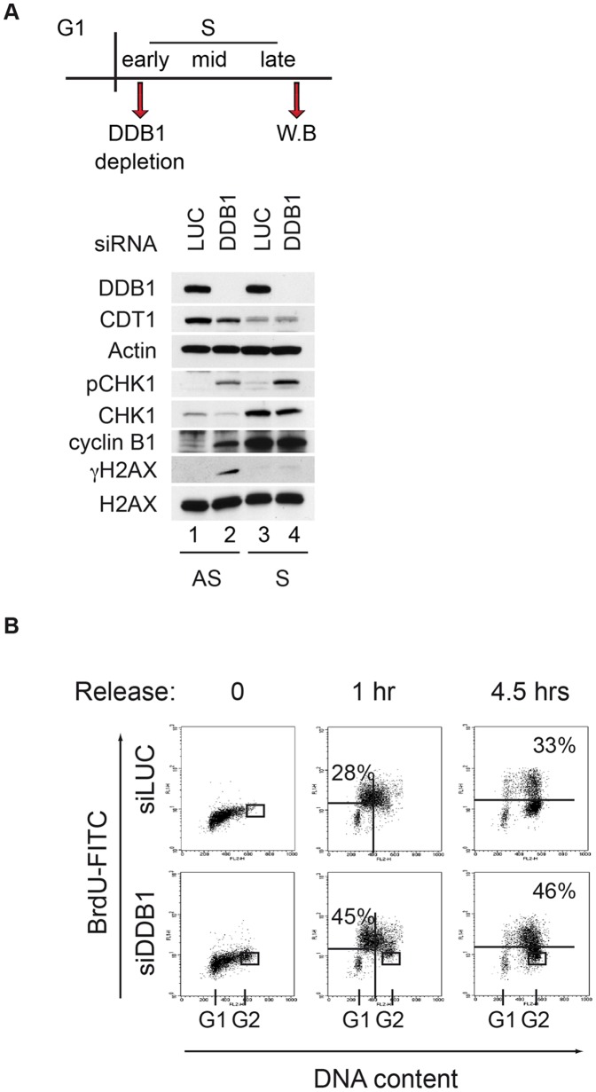
Figure 3. DDB1 depletion causes replication stress.
(A) Total protein extracts of HeLa cells depleted with the indicated siRNAs were fractionated by SDS-PAGE and immunoblotted with the indicated antibody. AS (asynchronous) indicates exponentially growing cell total protein extracts. S (synchronous) indicates total protein extracts from HeLa cells harvested 5 hours after releasing from a DTB. The experimental set up is summarized above the immunoblot. (B) HeLa cells were transfected with control or siDDB1 during DTB. Cells were labeled with BrdU and harvested at the indicated time points following DTB. Cells were immunostained with anti-BrdU antibody and DNA content was monitored by flow cytometry using propidium iodide staining. A representative FACS profile of three independent experiments with similar results is shown. Percentage of cells incorporating BrdU in early S-phase (1 hr) or late S-phase (4.5 hrs) was calculated. A minor population of permanently arrested G2 cells was detected in DDB1-depleted cells and was not considered in quantifications (delimitated by rectangle). G1 indicates cells with DNA content 2C; G2 indicates cells with DNA content 4C.

