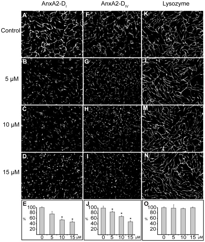
Figure 2. The effects of soluble AnxA2-DI, AnxA2-DIV and lysozyme on the formation of an in vitro capillary-like network.
Co-cultures of SMCs and GFP-expressing HUVECs were treated with 5–15 µM AnxA2-DI (B–D), AnxA2-DIV (G–I), lysozyme (L–N) at 2 h after seeding. Panels A, F and K show the corresponding controls with untreated cells. After 72 h incubation, images were taken at 10× magnification. The tube total length was analysed (E, J and O) and expressed as percentage relative to the corresponding untreated EC controls (100%) (A, F and K, respectively), as described for Figure 1. Results (E, J and O) are the mean ± SEM of 3 independent experiments each. Statistical significance was determined by the two-tailed Student's t-test (*P<0.05).

