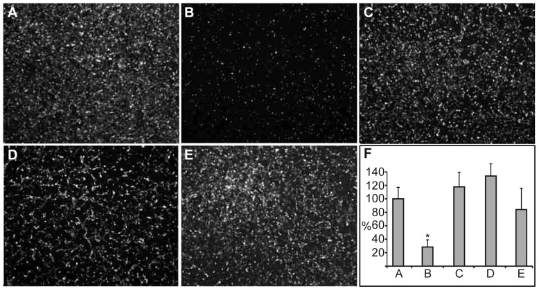
Figure 6. AnxA2-DI and AnxA2-DIV do not inhibit the migration of GFP-expressing HUVECs through a fibrin clot in a trans-well assay.
Images of GFP-expressing HUVECs migrated to the backside of a filter with a fibrin clot on its upper side, onto which the HUVECs were seeded. Control HUVECs (A); HUVECs incubated with 100 nM PTK787 (B), 15 µM lysozyme (C), AnxA2-DI (D), or AnxA2-DIV (E), which were present in both the upper and lower chambers. Migration is expressed as percentage relative to the untreated EC control (100%) (A). Results (F) are the mean ± SEM of 3 independent experiments each. Statistical significance was determined by the two-tailed Student's t-test (*P<0.05).

