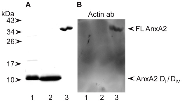
Figure 7. AnxA2-DI and AnxA2-DIV do not bind to α-actin.

∼10 µg of AnxA2-DI (∼55 µM) (lane 1), AnxA2-DIV (∼55 µM) (lane 2) and 5 µg of AnxA2 (∼6 µM) (lane 3) were separated by 15% SDS-PAGE (A and B) and transferred to a nitrocellulose membrane (B). Proteins were visualised by Coomassie Brilliant Blue staining (A). Far-Western (B); after denaturation and renaturation as described in Methods, the proteins were subjected to an actin overlay assay by incubation ON with 10 µg/ml α-actin and subsequent detection of bound actin by monoclonal actin antibodies. The positions of full-length (FL) AnxA2, and the domains I and IV of AnxA2 are indicated by arrowheads to the right. Selected standards are indicated by arrowheads to the left.
