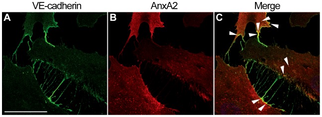
Figure 8. AnxA2 and VE-cadherin co-localise in endosome- and filopodia-like structures in sub-confluent HUVECs grown as a monolayer.

The cells were fixed in 3% paraformaldehyde and permeabilised with 0.05% Triton X-100 in PBS before further processing for dual label immunofluorescence using antibodies directed against endogenous VE-cadherin (A) and AnxA2 (B). (C) shows the merged image. Several sites of VE-cadherin and AnxA2 co-localisation are indicated by arrowheads. Bar, 40 µm.
