Abstract
Background:
Different surgical procedures that reduce orthodontic treatment time have been recommended. The aim of this study was to evaluate the effectiveness of a new less invasive surgical technique (called flapless bur decortications) for accelerating orthodontic tooth movement.
Materials and Methods:
This study was designed as a split-mouth study. The left and right maxillary first premolars of five dogs were extracted. An A-NiTi closed coil spring and an absolute anchorage was used for premolar protraction in both sides. In each dog, decortications were performed for one side and the other side was used as the control group. The distance between canine and second premolar was measured and the sites of decortication were evaluated histologically. The data was analyzed by paired samples t-test and multivariate analysis of two-way repeated measures ANOVA.
Results:
The study teeth moved more than their controls in the first month and less than their controls in the third month (P < 0.05). The total difference between study and control movements was not significant (P < 0.05).
Conclusion:
(1) Corticotomy facilitated orthodontic tooth movement is achievable with flapless bur decortication technique. (2) Velocity of tooth movement decreases in later stages of treatment due to maturation of newly formed bone at decortication sites.
Keywords: Accelerated orthodontic tooth movement, corticotomy, decorication
INTRODUCTION
The prolonged duration of orthodontic treatments is probably the chief complaint of both clinicians and patients. To reduce the treatment period, a thorough understanding of influencing factors on the orthodontic tooth movement is crucial. Recent cellular, molecular, and tissue engineering studies affirmed that mechanical stimuli are the most important, though not the exclusive inducing factor for orthodontic tooth movement. Accordingly, other stimuli such as surgical,[1] laser,[2] electromagnetic,[3,4] pharmaceutical[5,6] , and ultrasound[7] have been used along with mechanical stimuli to accelerate tooth movements.
Resorption and formation of cortical bone are the two basic phenomena which make orthodontic tooth movement possible.[8] All of the mentioned stimuli affect orthodontic movement by modulating the primary biological phenomena responsible for tooth movement.
These stimuli are based on regional acceleration phenomenon (RAP).[9] Frost demonstrated that if there is sufficient noxious stimulus, a series of physiologic healing responses, termed as RAP, can be induced, which are characterized with accelerated reorganizing activity in osseous and soft tissues.[9]
Surgical injuries are potent stimuli in the induction of RAP. Based on this rationale, different dentoalveolar supplemental surgical procedures have been recommended. Liou and Huang[10] proposed periodontal ligament distraction and Iseri et al.[11] suggested dentoalveolar distraction to perform rapid canine retraction. In addition, many clinicians like Wilcko et al.[12–14] Suya,[8] Kole et al.,[15] and Badr et al.[16] have attempted different methods of decortications; they have all confirmed the effectiveness of these techniques. There are also some recent animal studies that investigated the effects of corticotomy facilitated orthodontics. For example, Ren et al.,[17] Lino et al.[18] , and Mostafa et al.[19] all reported that their methods of decortication after reflecting surgical flaps were effective in accelerating tooth movement compared to control.
Despite the repeated confirmation of the effectiveness of corticotomy-facilitated orthodontics through numerous case reports and animal studies, this technique has not become popular clinically due to its invasiveness. Consequently, research into other less invasive procedures that may facilitate orthodontic treatment is required.
Kim et al.[1,7] introduced corticision technique that is a surgical method with minimal intervention; it is reportedly effective in accelerating tooth movement through induction of RAP. Despite its minimal intervention, four vertical incisions are required in this technique; and transecting the mucogingival line could lead to postoperative complications.
To reduce the treatment time with minimal invasiveness, we introduced a new technique called flapless bur decortication. This technique contains no flap retraction. Instead, small holes are made along the buccal bone of the tooth that requires orthodontic movement using a fine surgical fissure bur. Since the holes are located in the attached gingiva using lowspeed/high-torque rotary instrument, minimal complications are expected. The purpose of this study was to evaluate the effectiveness of this new technique in accelerating orthodontic tooth movement and its potential complications.
MATERIALS AND METHODS
Ten premolar teeth of five adult German male dogs, weighting 20-25 kg with the age range of 18-24 months were used in this study. Animals were supervised in the hospital of small animals (Tehran University, Tehran, Iran) for one week prior to the study. Their health status was evaluated by an experienced veterinary surgeon according to the animal welfare regulations. The animals were caged individually and fed with dog food. Animals were surgically preanesthetized with Ketamine (10 mg/kg, IM) and 2% Xylazine (5 mg/kg) and 5% Thiopental natrium (10 mg, IV). Before the operation, the oral cavity was irrigated with 0.2 % povidine iodine and 0.02 % chlorhexidine mouthwash, respectively.
The total of ten left and right maxillary first premolars were extracted [Figure 1].
Figure 1.
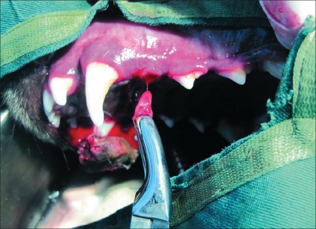
First premolar tooth was extracted following local anesthesia
Miniscrews (Jeil Medical Corporation, Korea. 8-mm length 1.4 mm diameter, self-drilling) were placed mesial to left and right upper canines. A small groove was created in the most gingival part of clinical crown of right and left upper second premolars using a porcelain disk. A10-mm A-NiTi closed coil spring (Orthotechnology corporation, USA) was used for premolars protraction. The two ends of ligature wire were ligated; one around the cervical groove of second premolar and one to the miniscrew head using steel ligature wire (3 M-Unitek, 0.01 inch). The ligature wires were stabilized using Nomix adhesive (3 M Unitek Corporation, USA) [Figure 2].
Figure 2.
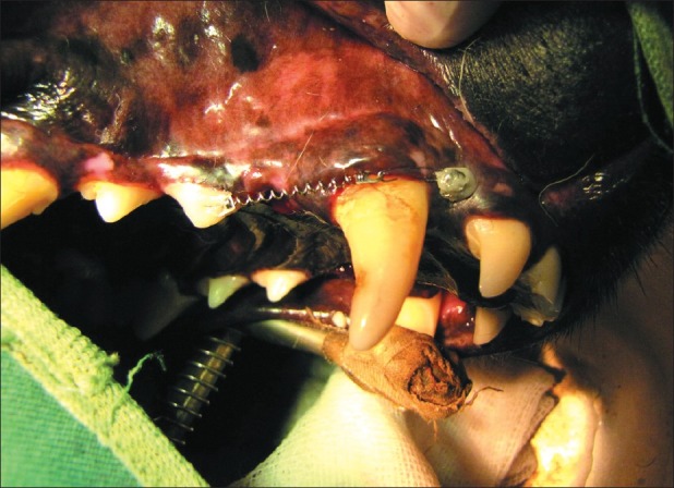
The force system used for second premolar protraction; the ligated wires were stabilized using Nomix adhesive
A 150-gr orthodontic force was initially exerted for protraction of the second premolar.
Decortication was performed for one side of each arch and the other side was used as the control.
During the process of decortication, small holes, without gingival flaps, were created through mesial and distal attached gingiva of each second premolar tooth. The holes were made on each side using a pointed tungsten carbide fissure bur (The contouring fissure bur C1, Technicare Dental Supplies Ltd. GB). Also, other holes were made on the buccal cortical table of the extracted tooth. The total number of these holes was twenty five [Figure 3].
Figure 3.
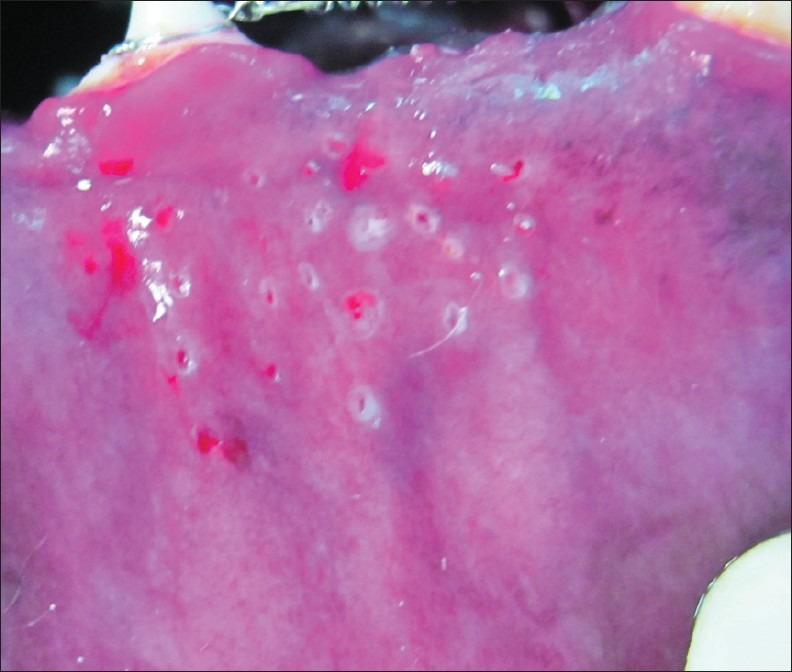
Small holes made mesial and distal to the second premolars and on the buccal table of extracted first premolar
The holes (all with 2-mm depth) were prepared using high torque, low speed surgical handpiece along with copious irrigation.
Antibiotic (Penicillin 633, od), analgesics (Mephenamic Acid 250 mg, bd/bid and tramadol 100 mg, bd/bid) were prescribed for 5 days post-operatively.
The decortication process was repeated at the end of the first and second month.
Two 1-mm depth pointed holes were made on the buccal surface of canines and second premolars with the same tungsten carbide fissure bur. The inter-points distances were recorded at the beginning of the experiment and one, two and three months thereafter by a digital caliper. Measurements were made to the nearest 0.01 mm [Figure 4].
Figure 4.
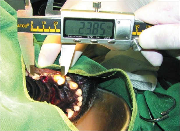
The inter-points distances were measured with digital caliper (accuracy = 0.01 mm)
Two measurements were taken for each side by two independent examiners and the average was considered as the data.
The condition of each miniscrew, attached gingiva, operation site, and the teeth were checked monthly. Also, the coil springs were reactivated in the case of diminished forces; oral cavity was irrigated with 0.02% chlorhexidine. The decortication sites were evaluated for postsurgical complications such as fistula formation, ulceration, hematoma, and lack of proper healing.
The dogs were killed at the end of the study period for histologic procedure. The experiment was performed according to the Ethics Committee of Shahid Beheshti University of Medical Sciences. Specimens including decortication sites were dissected. The specimens were decalcified in 10% formic acid and after preparation of paraffin blocks were sectioned mesiodistally (3-5 μm thick) and stained with hematoxylin and eosin technique.
The amounts of tooth movements at the both sides during the first, second, and third months were calculated. Data were analyzed using SPSS (version 16.0).
Data analysis
This study was designed as a split-mouth study. Descriptive statistics (mean, standard deviation) for velocity of monthly movement of second premolars in each group and accumulated distance of second premolar movement after each month were used in all groups.
Based on normal distribution of the data (Shapiro-Wilk test), paired samples t-test was used for intergroup comparisons of mean tooth movements at each time. For intergroup and intragroup comparison of tooth movement by time period (i.e., monthly changes), we used the multivariate analysis of two-way repeated measures ANOVA. Because the sphericity was not assumed according to Mauchly's test of sphericity, the Greenhouse-Greisser correction was used for intergroup comparison of tooth movements by time. A value of P < 0.05 was considered statistically significant.
RESULTS
The monthly movement and accumulated distance values for second premolars in study and control groups are shown in Table 1.
Table 1.
Comparison of the velocity and accumulated amount of second premolars movements in the study and control groups
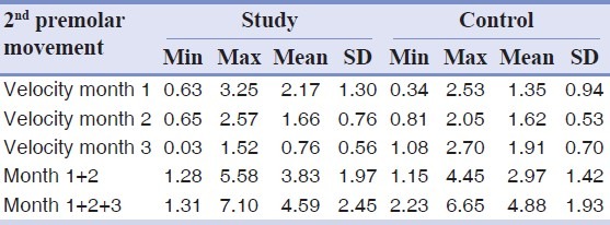
Accordingly, during the first month, the movement of second premolars in the decortications side was 0.82 (±0.56) mm greater than controls and this difference was statistically significant (P = 0.031). This difference was 0.04 (±0.0.40) mm during the second month; not statistically significant (P = 0.826).
The difference between tooth movements during the third month of the experiment was 1.15 (±0.58) mm. During this time, the second premolars in the decortications side moved less than the control side and this difference was significant (P = 0.012).
Considering the accumulated distance of tooth movement, the difference between accumulated tooth movement after two months was 0.86 (±0.75) mm and this difference was not significant (P = 0.063). The difference between total amounts of tooth movement after three months between study and control groups was 0.29 (±0.0.93) mm and this difference was not significant (P = 0.527) [Table 1].
The results of this survey revealed that the differences between the amount of second premolar movement during the first, second and third months of the study were not significant for the control group (P = 0.240). In contrast, the velocity of this movement in the study group was significantly affected by the time (P = 0.037, Greenhouse-Geisser correction).
The general patterns of monthly velocity of tooth movement in study and control groups were totally different [Figure 5].
Figure 5.
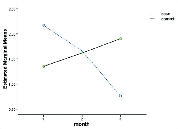
Comparative patterns of movement velocity related to second premolars during postoperative three months
In the study group, the highest velocity of tooth movement was observed during the first month, followed by the second and third month in respect. This demonstrated a descending pattern for movement velocity in study group. Contrarily, the related pattern for controls was increasing during 1st-3rd month, but not significantly different.
Considering the postsurgical complications, there was no findings of hematomas, ulcerations or oronasal fistulas during the study period. However, despite complete healing of the oral mucosa two weeks postsurgically, clinical appearance of decortications site was distinguishable.
The sites of decortication had two different histologic views [Figures 6 a–b].
Figure 6(a,b).
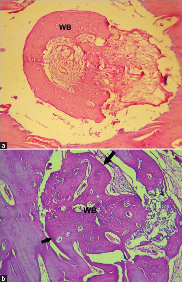
Microphotographs of different decortication sites; (a), a site filled with woven bone with large marrow spaces; (b), More compact lamellar bone (arrows) has started to form from periphery, WB, woven bone(H and E staining, ×200)
Some of these sites were filled with the woven bone with large marrow spaces. The other sites were filled with more compact lamellar bone.
DISCUSSION
The results of the present study indicated that in spite of effectiveness during the first month, flapless bur decortication technique had an inhibitory effect on orthodontic tooth movements in later stages. Limited or less than enough area of decortications probably played an important role in limited acceleration of tooth movement during the first month.
It can been proposed that the greater the amount of surgical intervention (decortications), the greater the availability of biologic materials necessary for accelerating orthodontic tooth movement and the faster the velocity of tooth movement. Frost[20] observed a direct correlation between severity of surgical insult and intensity of regional acceleration phenomenon. Mostafa et al.[19] reported a twice amount of accelerated tooth movement in study group compared to control group during the first month. In our study, 1.6 fold tooth movement in study group was found. This finding suggests that reducing the area of decortication negatively affects accelerated orthodontic tooth movement. In practice, few patients accept the risks of very wide area of decortication despite its effectiveness in reducing treatment time. Then, the following questions will arise: 1 - What is the minimum amount of this intervention to successfully accelerate tooth movement? 2 - Does decortications in left side contribute to accelerating tooth movement in the right side? 3 - Does (would) local decortication around the teeth, which are subjected to orthodontic movement, effectively accelerate tooth movement? Another indistinct issue is the location of decortication to be either buccal and lingual or buccal.
During the third month, the oldest sites of decortication went through the process of bone lamellation and maturation [Figures 6b and 7] and adversely affected tooth movement. Biologic materials recruited because of decortication not only increased bone turn over, but also effectively contributed to accelerating bone maturation. As a result, the bone formed distal to the mesial root of second premolars on the experiment side was of the more mature lamellar bone in comparison with the relatively immature bone with wider marrow spaces on the control side. Mostafa et al.[19] suggested this difference in quality of the newly formed bone as a reason for the reduced relapse tendency in the experiment side. This difference at the same time retarded tooth movement after the mesial side of distal root reached this bone in experiment side. Recent clinical as well as animal studies support the declining pattern of tooth movement in study group at later stages of the treatment.[16,21]
Figure 7.
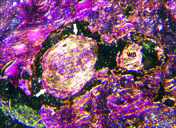
Phase contrast microphotography of two adjacent decortication sites; (a), woven bone (WB); (b), lamellar bone in the periphery (arrow) (×40)
During the second month the effect of decotication was neutralized by the resistance to tooth movement provided by the process of bone maturation.
The most important limitation in our technique was limited area available for decortication. For more extended decortication in larger areas, there was the risk of nasal floor perforation due to the vicinity of this structure with the second premolar teeth in dogs. The available area of attached gingiva was the second limiting factor because decortication in the alveolar mucosa had the risk of tissue rupturing and unfavorable bleeding.
In regard to using slow-speed hand-piece with copious irrigation and also the attached gingival as the holes’ site, minimal risk of complications was expected. That was achieved through the results of our study since no postoperative complications were observed during the three month observation of our experiment. If this technique would be tested in humans, the method of creating small holes between the roots of teeth could be modified by using hand instruments; like the ones used for mini-implants, to improve safety of the method and decreasing the probability of complications.
CONCLUSIONS
Corticotomy facilitated orthodontic tooth movement is achievable with flapless bur decortication technique.
Velocity of tooth movement decreases in later stages of treatment due to maturation of newly formed bone at decortication sites.
As the timing of physiological events responsible for tooth movement in animal models differs from humans, future clinical trials with similar basis are recommended for determining timing of these physiological events in humans.
Footnotes
Source of Support: Nil
Conflict of Interest: None declared
REFERENCES
- 1.Kim SJ, Park YG, Kang SG. Effects of Corticision on paradental remodeling in orthodontic tooth movement. Angle Orthod. 2009;79:284–91. doi: 10.2319/020308-60.1. [DOI] [PubMed] [Google Scholar]
- 2.Kawasaki K, Shimizu N. Effects of low-energy laser irradiation on bone remodeling during experimental tooth movement in rats. Lasers Surg Med. 2000;26:282–91. doi: 10.1002/(sici)1096-9101(2000)26:3<282::aid-lsm6>3.0.co;2-x. [DOI] [PubMed] [Google Scholar]
- 3.Davidovitch Z, Finkelson MD, Steigman S, Shanfeld JL, Montgomery PC, Korostoff E. Electric currents, bone remodeling, and orthodontic tooth movement. II. Increase in rate of tooth movement and periodontal cyclic nucleotid levels by combined force and electric current. Am J Orthod. 1980;77:33–47. doi: 10.1016/0002-9416(80)90222-5. [DOI] [PubMed] [Google Scholar]
- 4.Stark TM, Sinclair PM. Effect of pulsed electromagnetic fields on orthodontic tooth movement. Am J Orthod Dentofacial Orthop. 1987;91:91–104. doi: 10.1016/0889-5406(87)90465-3. [DOI] [PubMed] [Google Scholar]
- 5.Leiker BJ, Nanda RS, Currier GF, Howes RI, Sinha PK. The effects of exogenous prostaglandins on orthodontic tooth movement in rats. Am J Orthod Dentofacial Orthop. 1995;108:380–8. doi: 10.1016/s0889-5406(95)70035-8. [DOI] [PubMed] [Google Scholar]
- 6.Igarashi K, Mitani H, Adachi H, Shinoda H. Anchorage and retentive effects of a bisphosphonate (AHBuBP) on tooth movements in rats. Am J Orthod Dentofacial Orthop. 1994;106:279–89. doi: 10.1016/S0889-5406(94)70048-6. [DOI] [PubMed] [Google Scholar]
- 7.Kim SJ, Moon SU, Kang SG, Park YG. Effects of low-level laser therapy after Corticision on tooth movement and paradental remodeling. Lasers Surg Med. 2009;41:524–33. doi: 10.1002/lsm.20792. [DOI] [PubMed] [Google Scholar]
- 8.Suya H. Corticotomy in orthodontics. In: Hosl E, Baldauf A, editors. Mechanical and biological basics in orthodontic therapy. Heidelberg Germany: Huthig Buch Verlag; 1991. pp. 207–26. [Google Scholar]
- 9.Frost HA. The regional acceleratory phenomena: A review. Henry Ford Hosp Med J. 1983;31:3–9. [PubMed] [Google Scholar]
- 10.Liou EJW, Huang CS. Rapid canine retraction through distraction of the periodontal ligament. Am J Orthod Dentofacial Orthop. 1998;114:372–82. doi: 10.1016/s0889-5406(98)70181-7. [DOI] [PubMed] [Google Scholar]
- 11.Iseri H, Kisnisci R, Bzizi N, Tuz H. Rapid canine retraction and orthodontic treatment with dentoalveolar distraction osteogenesis. Am J Orthod Dentofacial Orthop. 2005;127:533–41. doi: 10.1016/j.ajodo.2004.01.022. [DOI] [PubMed] [Google Scholar]
- 12.Wilcko WM, Wilcko T, Bouquot JE, Ferguson DJ. Rapid orthodontics with alveolar reshaping: Two case reports of decrowding. Int J Periodontics Restorative Dent. 2001;21:9–19. [PubMed] [Google Scholar]
- 13.Wilcko WM, Ferguson DJ, Bouquot JE, Wilcko MT. Rapid orthodontic decrowding with alveolar augmentation: Case report. World J Orthod. 2003;4:197–205. [Google Scholar]
- 14.Wilcko MT, Wilcko WM, Nabil F. Bissada. An evidencebased analysis of periodontally accelerated orthodontic and osteogenic techniques: A synthesis of scientific perspectives. Semin Orthod. 2008;14:305–16. [Google Scholar]
- 15.Kole H. Surgical operation on the alveolar ridge to correct occlusal abnormalities. Oral Surg Oral Med Oral Pathol Oral Radiol Endod. 1959;12:515–29. doi: 10.1016/0030-4220(59)90153-7. [DOI] [PubMed] [Google Scholar]
- 16.Aboul-Ela SM, El-Beialy AR, El-Sayed KM, Selim EM, El-Mangoury NH, Mostafa YA. Miniscrew implant-supported maxillary canine retraction with and without corticotomyfacilitated orthodontics. Am J Orthod Dentofacial Orthop. 2011;139:252–9. doi: 10.1016/j.ajodo.2009.04.028. [DOI] [PubMed] [Google Scholar]
- 17.Ren A, Lv T, Zhao B, Chen Y, Bai D. Rapid orthodontic tooth movement aided by alveolar surgery in beagles. Am J Orthod Dentofacial Orthop. 2007;131:160.e1–10. doi: 10.1016/j.ajodo.2006.05.029. [DOI] [PubMed] [Google Scholar]
- 18.Iino S, Sakoda S, Ito G, Nishimori T, Ikeda T, Miyawaki S. Acceleration of orthodontic tooth movement by alveolar corticotomy in the dog. Am J Orthod Dentofacial Orthop. 2007;131:448.e1–8. doi: 10.1016/j.ajodo.2006.08.014. [DOI] [PubMed] [Google Scholar]
- 19.Mostafa YA, Fayed MM, Mehanni S, ElBokle NN, Heider AM. Comparison of corticotomy-facilitated vs standard tooth movement techniques in dogs with miniscrews as anchor units. Am J Orthod Dentofacial Orthop. 2009;136:570–7. doi: 10.1016/j.ajodo.2007.10.052. [DOI] [PubMed] [Google Scholar]
- 20.Frost HM. The regional accelerated phenomenon. Orthop Clin N Am. 1981;12:725–6. [Google Scholar]
- 21.Baloul SS, Gerstenfeld LC, Morgan EF, Carvalho RS, van Dyke TE, Kantarci A. Mechanism of action and morphologic changes in the alveolar bone in response to selective alveolar decortication-facilitated tooth movement. Am J Orthod Dentofacial Orthop. 2011;139:S83–101. doi: 10.1016/j.ajodo.2010.09.026. [DOI] [PubMed] [Google Scholar]


