Figure 2.

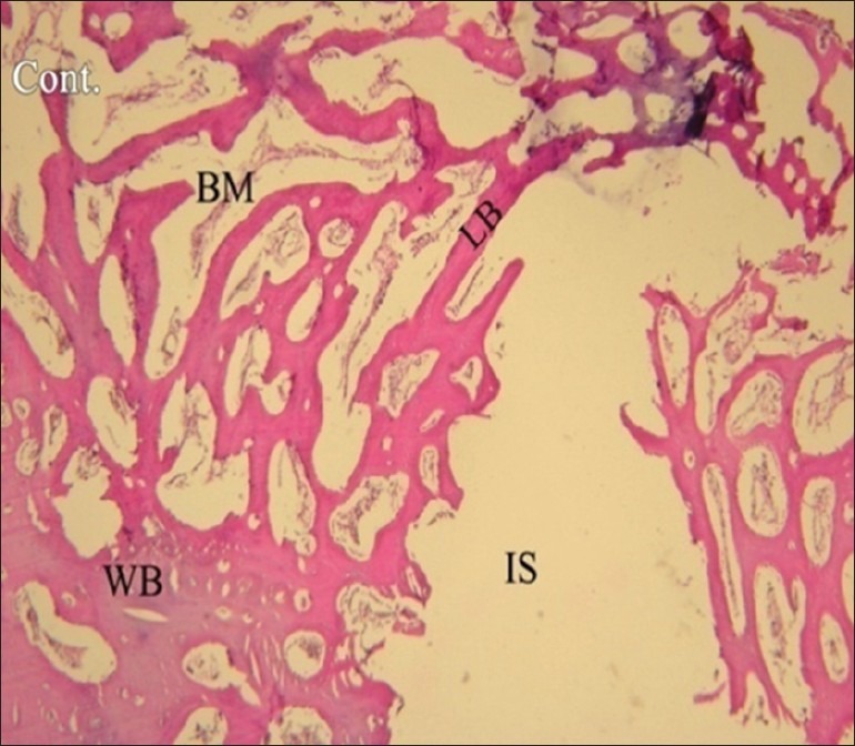
Histological image taken under a light microscope in control group in the fourth week. (H and E, magnification ×40). IS: Implant surface, WB: Woven bone, LB: Lamellar bone, BM: Bone marrow

Histological image taken under a light microscope in control group in the fourth week. (H and E, magnification ×40). IS: Implant surface, WB: Woven bone, LB: Lamellar bone, BM: Bone marrow