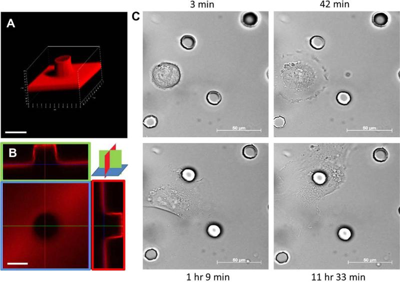
Fig. 1.
Confocal images of post and cell interaction: (A) 3D image of a post 15 μm high, and 15 μm diameter with laminin coating. Scale bar, 20 μm. (B) Uniform laminin staining on flat base and sides of the post seen by confocal microscopy. Scale bar, 20 μm. (C) Time lapse images show initial migration and preferential adhesion of a cell to a post during the following 12 h. Scale bar, 50 μm.

