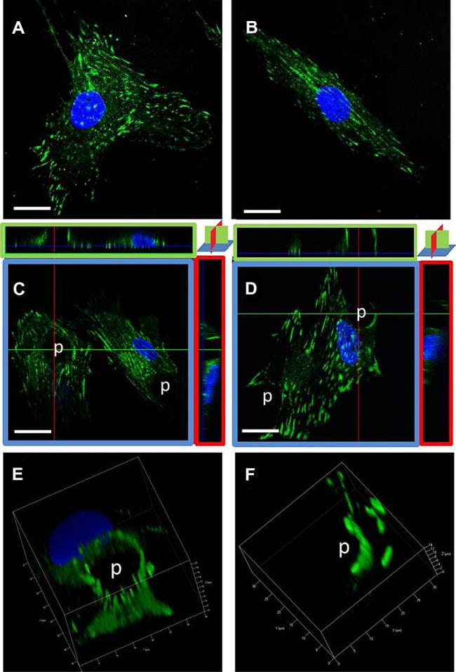
Fig. 3.
Focal adhesions redistributed with microtopography and strain. Paxillin staining of hMSCs after two days with or without straining at 1 Hz and 10% strain seen in 3D with confocal microscopy. (A) flat, (B) flat-strain, (C) post confocal orthogonal views, (D) post-strain orthogonal views. Volumetric renditions of 3D for (E) post and (F) post-strain. (C) Paxillin surrounds the post and goes up its full height. (D) Paxillin forms a semicircular ring up the side of the post, and extends from post to post on the lower surface of the cell. (E, F) 3D reconstructions show punctate paxillin on the sides of the post. Paxillin (green) and DAPI (blue). Scale bar, 20 μm. (For interpretation of the references to color in this figure legend, the reader is referred to the web version of this article.)

