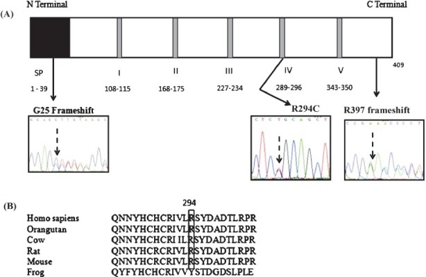
Fig.

(A). Schematic representation of the human Neu1 protein with the relative positions of the three identified mutations marked by black arrows. The signal peptide region is indicated by a black box marked SP and the five conserved Asp box motifs are denoted by gray bands; the amino acids included in each are mentioned. The chromatograms show the sequences at the mutated sites. (B). Clustal 2.1 multiple sequence alignment of the human Neu1 protein sequence against the Neu1 protein sequences of other species shows that R (arginine) is conserved at the 294th position in other mammalian species also
