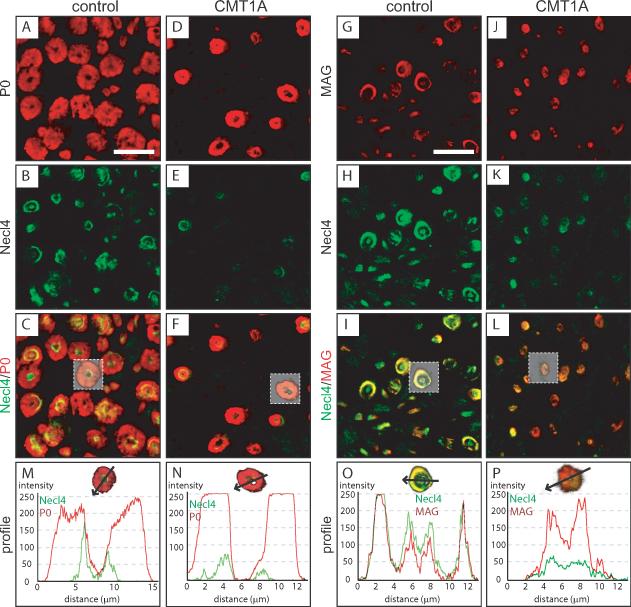
Figure 2. Altered periaxonal localization of MAG and Necl4 in CMT1A patients.
Immunofluorescent confocal colocalization of transversal sections from sural nerves of control autopsies (A–C, G–I) and CMT1A biopsies (D–F, J–L) for Necl4 (B, E, H, K) and either P0 (A, D) or MAG (G, J). Immunofluorescence for Necl4 was strongly decreased in nerve tissue from CMT1A patients (E, K, N, P). MAG and Necl4 were colocalized at periaxonal regions in control as well as in CMT1A patients (I, L, O, P). Analysis of fiber profiles revealed an altered ratio of immunolabeling for MAG and Necl4 at the periaxonal region in CMT1A patients (P). A reduction of Schmidt-Lanterman incisures in CMT1A is observed, which is due to the loss of large caliber fibers in CMT1A, which have many more Schmidt-Lanterman incisures than small caliber fibers. Scale bar: 20 μm.

