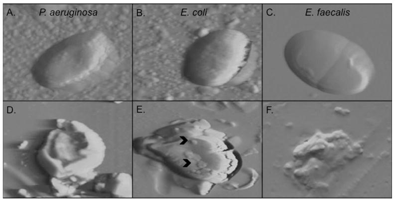
Figure 7. RNase 7 disrupts the microbial membrane at low micromolar concentrations.
Atomic force microscopy demonstrates that 2.5 μM of RNase 7 disrupts the structural integrity of P. aeruginosa, E. coli, and E. faecalis after 45 minutes of exposure. A–C: Images of untreated uropathogens revealed intact cell morphology with a relatively smooth surface. D–F: uropathogens treated with RNase 7 showed loss of cellular integrity with membrane splitting and bleb formation (^). Each panel is 5 micron.

