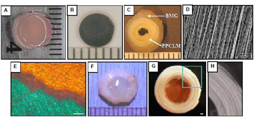
Figure 6.
Selected biomaterials for IVD repair. (A) Photocrosslinked carboxymethylcellulose hydrogel with encapsulated NP cells (B) Fibrin-Genipin adhesive for AF fissures (C) Biphasic scaffold for AF repair consisting of concentric sheets of poly(polycaprolactone triol malate) (PPCLM) surrounded by a ring of demineralized bone matrix gelatin (BMG) (Wan et al., 2008). (D) Scanning electron micrograph of an electrospun polycarbonate polyurethane nanofibrous scaffold for AF replacement (Yeganegi et al., 2010). (E) Polarized light micrograph of an electrospun poly(e-caprolactone) (PCL) nanofibrous scaffold oriented in opposing bilayers (+30°/−30°) seeded with mesenchymal stem cells which elaborate aligned intra-lamellar collagen that recapitulates the gross fiber orientation of the native AF (Nerukar et al., 2009). Scale: 200 µm (F) Composite whole disc equivalent comprised of NP cells encapsulated in an alginate hydrogel surrounded by a reinforced poly(glycolic acid) mesh seeded with AF cells (Mizuno et al., 2006). (G) Disc-like angle-ply structure constructed from PCL nanofibers oriented at +30°/−30° to mimic the AF with a central agarose hydrogel core serving as an NP analog (Nerukar et al., 2010). Scale: 1 mm. (H) Higher magnification view of AF region outlined in panel G. Published images reprinted with permission from Elsevier (C, D, F), Nature Publishing Group (E) and Wolters Kluwer (G, H).

