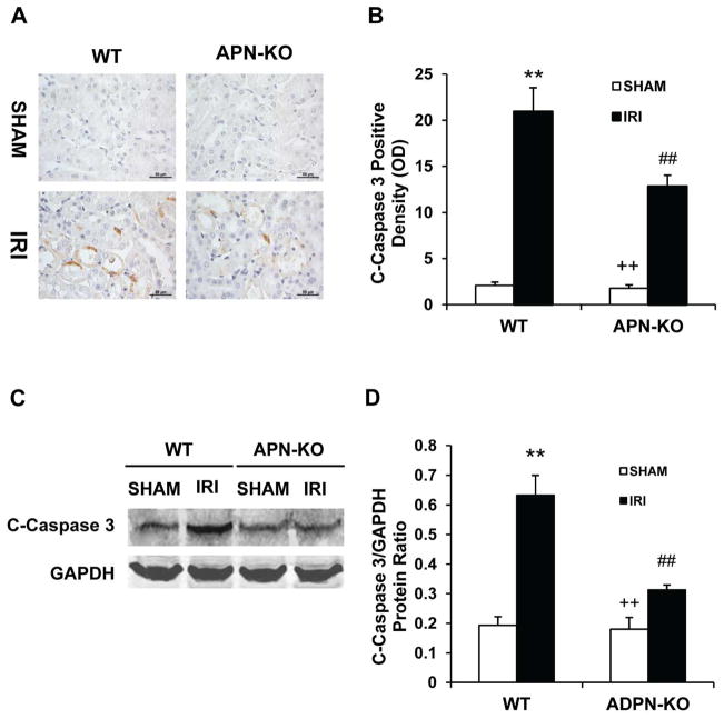
Figure 4. Adiponectin deficiency inhibits caspase 3 activation in tubular epithelial cells.
A. Representative photomicrographs of kidney sections stained for cleaved caspase 3 (brown) and counterstained with hematoxylin (blue) in WT and APN-KO mice after sham or IRI. (Original magnification: X400). B. Quantitative analysis of cleaved caspase 3 expression in kidneys of WT and APN-KO mice after sham or IRI. **P < 0.01 vs WT controls; ## P < 0.01 vs WT IRI, ++ P < 0.01 vs APN-KO IRI. n=5–6 in each group. C. Representative Western blots show cleaved caspase 3 protein expression in kidneys of WT and APN-KO mice after sham or IRI. D. Quantitative analysis of cleaved caspase 3 protein expression in kidneys of WT and APN-KO mice after sham or IRI. **P < 0.01 vs WT sham; ## P < 0.01 vs WT IRI, ++ P < 0.01 vs APN-KO IRI. n=5–6 in each group.

