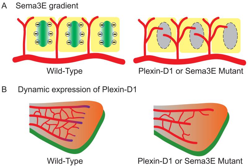
Fig. 1. Temporal and spatial control of ligand vs. receptor results in two different mechanisms governing vascular patterning.
(A) The repulsive gradient generated by Sema3E in the mouse somite determines the proper patterning of Plexin-D1-expressing intersomitic vessels [4]. During intersomitic vessel (red) development in the mouse embryo, Sema3E (gradient in green) is expressed in the caudal region of each somite (yellow), whereas Plexin-D1 is expressed in the adjacent intersomitic vessels (red) on the rostral region of each somite. The repulsive cues generated by the Sema3E gradient restrict vessel growth and branching in the intersomitic space. Mice lacking Sema3E or Plexin-D1 lose the repulsive gradient signals (grey oval), thereby allowing blood vessels to encroach on somites and display exuberant blood vessel growth in the entire somite and a loss of the normal segmented pattern.
(B) In the retina, dynamic regulation of Plexin-D1 level instead of a Sema3E gradient is crucial to establish properly patterned retinal vasculature [15, 16]. In contrast to Sema3E gradient in the intersomitic vessels, in the retinal vasculature (red), Plexin-D1 is selectively expressed in endothelial cells (purple gradient) at the front of sprouting blood vessels in response to the VEGF gradient (orange), whereas Sema3E (green) is evenly expressed in RGCs underneath the retinal vasculature. The dynamic regulation of Plexin-D1 by VEGF in the sprouting front cells modulates the ratio between tip and stalk cells via VEGF-induced feedback mechanism to ensure balanced vascular network formation. Therefore, loss of dynamic Plexin-D1 regulation in the Plexin-D1 or Sema3E mutant shows less-branched and uneven front vasculature (right side of B).

