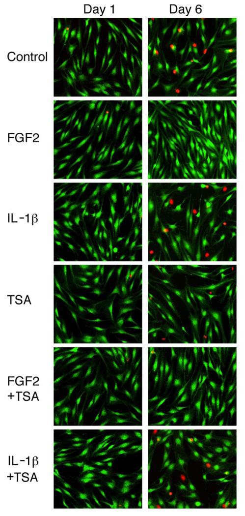
Figure 3.
Detection of live cells and dead cell nuclei in human articular chondrocyte cultures treated with FGF2, IL-1β, and TSA. Cells were cultured and treated as described in Figure 1. Green fluorescence indicates live cells and red fluorescence shows nuclei of dead cells.

