
Figure 2.
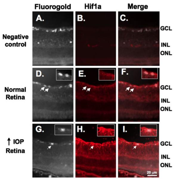
HIF-1α staining is not present in RGC. RGC were previously backlabeled with Fluoro-Gold (a, d, and g). Fluoro-Gold images were converted to gray scale to optimize visualization to determine colocalization with HIF-1α. b No nonspecific staining was observed when sections were incubated with secondary antibody alone. Subtle HIF-1α staining was observed in the inner layers of the normal retina (e) and was increased under conditions of high IOP (h). HIF-1α staining (red) did not colocalize with the RGC marker Fluoro-Gold (white) in the normal retina (f) or in the retina under conditions of high IOP (i). Insets are higher magnification views of the RGC indicated by arrows