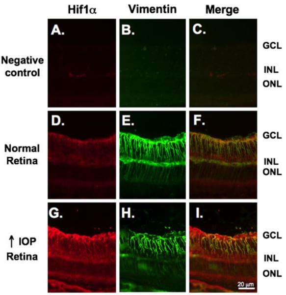
Figure 3.

HIF-1α expression is increased in Müller glia in the glaucomatous retina. Sections were double stained with an anti-HIF-1α antibody (red) followed by staining with an antivimentin antibody (green). a, b No nonspecific staining was observed when sections were incubated with their respective secondary antibodies alone. As shown in the same sections in Fig. 2, there is subtle HIF-1α staining in the inner layers of the normal retina (d) that increase under conditions of high IOP (g). d Low levels of HIF-1α staining colocalized (yellow) with the Müller glia marker vimentin under conditions of normal IOP. i Elevated HIF-1α expression was observed primarily in Müller glia as the staining colocalized with vimentin (yellow)
