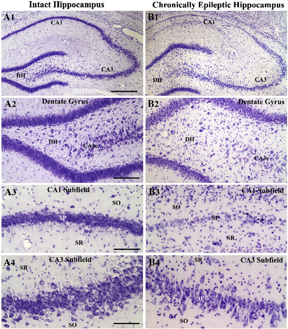
Fig. 1.
Cytoarchitecture of the hippocampus in an intact control rat (A1) and a chronically epileptic rat at 6 months post-status epilepticus (B1). A2–B4 illustrate magnified views of regions of dentate hilus (A2, B2), CA1 subfield (A3, B3) and CA3 subfield (A4 and B4) from A1 and B1. Note that, in comparison to the control hippocampus, the hippocampus of a chronically epileptic rat exhibits considerable loss of neurons in the dentate hilus (DH) and CA1 pyramidal cell layer and moderate loss of neurons in the CA3 pyramidal cell layer. DG, dentate gyrus; DH, dentate hilus; SO, stratum oriens; SP, stratum pyramidale; SR, stratum radiatum. Scale bar, A1 and B1=500 µm; A2 and B2=100 µm; A3, B3, A4, B4=50 µm.

