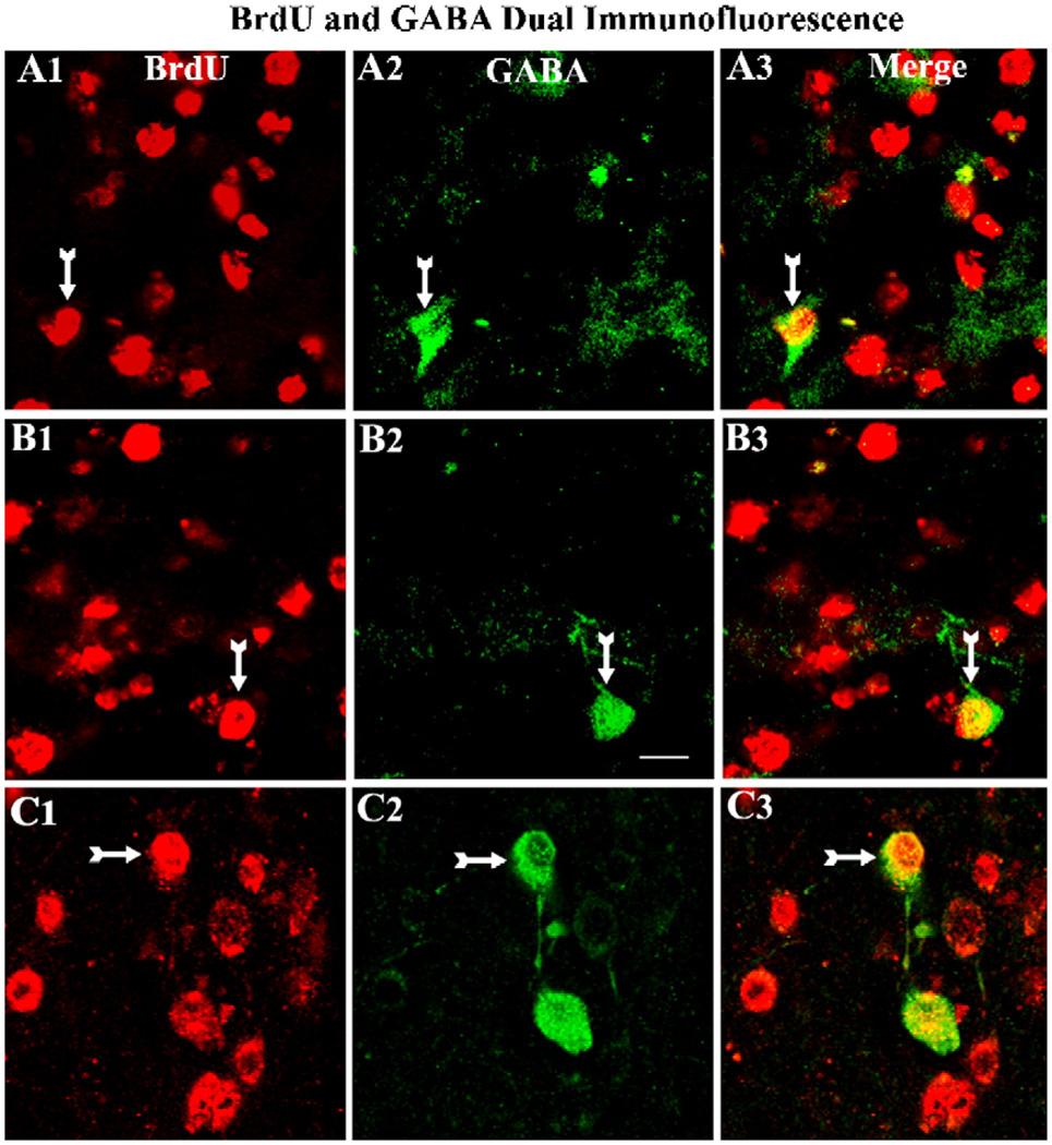
Fig. 6.
Differentiation of grafted cells into GABA-ergic interneurons. A1–C3 illustrate confocal images of grafted neurons that are positive for 5′-bromodeoxyuridine (BrdU) and GABA (arrows) in standard hippocampal fetal cell (HFC) grafts (A1–A3), HFC grafts treated with BDNF, NT-3 and caspase inhibitor (B1–B3) and HFC grafts treated with FGF-2 and caspase inhibitor (C1–C3). Note that only smaller fractions of transplanted cells differentiate into GABA-ergic interneurons in all groups. Scale bar, 10 µm.

