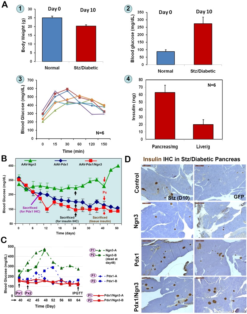
Figure 2.
Reversal of Stz/diabetes by liver-IPCs reprogrammed by AAV-PTFs. A. Baselines of Stz/diabetic NOD/SCID mice. NOD/SCID mice (10-12 weeks, n=6) were treated daily with Stz for five days and blood glucose levels and body weight were monitored every other day until the onset of diabetes (two measurements 24 h apart). Mice were sacrificed at glucose levels between 250 to 350 mg/dL and IPGTT was performed before the scarification. Tissue insulin was extracted from the pancreata and livers and quantified by ELISA. 1. Body weights of normal and diabetic NOD-SCID mice; 2. Blood glucose levels; 3. IPGTT; 4. Tissue insulin content of the liver and pancreas. Baseline information (1-3) was obtained just before the mice were sacrificed. B. Effects of AAV-mediated expression of PTFs on blood glucose levels. Stz-induced diabetic (>250 mg/dL) mice were injected via the portal vein with various AAV viruses (108 vp/mouse) expressing GFP, Ngn3, Pdx1, or a combination of Pdx1/Ngn3. Blood glucose levels were measured. Triangles, mice receiving AAV-Ngn3 (n=4); diamonds, mice receiving AAV-Pdx1 (n=8); squares, mice receiving both vectors (n=8). At day 24 post-injection, at least one mouse from each group was sacrificed for IHC (black arrows). At day 30, three mice from the AAV-GFP group (data not shown here) and AAV-Pdx1/Ngn3 groups were killed for tissue insulin measurements (brown arrow). Around day 40, two mice from each group underwent subtotal Px (red arrows). C. Changes of blood glucose levels following subtotal Px. To investigate a role of pancreatic beta-cells in normalizing the blood glucose levels, subtotal Px was performed on two mice from each group and changes of blood glucose levels were recorded (P1 and P2). Red lines represent mice receiving Pdx1/Ngn3; Blue lines, mice receiving Pdx1; and green lines, mice receiving Ngn3. One mouse in Ngn3 group (P2) died five days post surgery. D. Insulin IHC in Stz/Diabetic Pancreas. Formalin-fixed and paraffin embedded pancreatic sections were incubated with anti-insulin antibodies and visualized by DA B . Control (day 10) tissue was obtained after the first dose of Stz. The area shown was the only region containing islets in the entire pancreas. The remaining photographs are representative sections from Stz-induced diabetic mice at day-24 post-treatment via the portal vein with AAV-GFP, AAV-Ngn3, AAV-Pdx1, and AAV-Pdx1/Ngn3.

