
Figure 5.
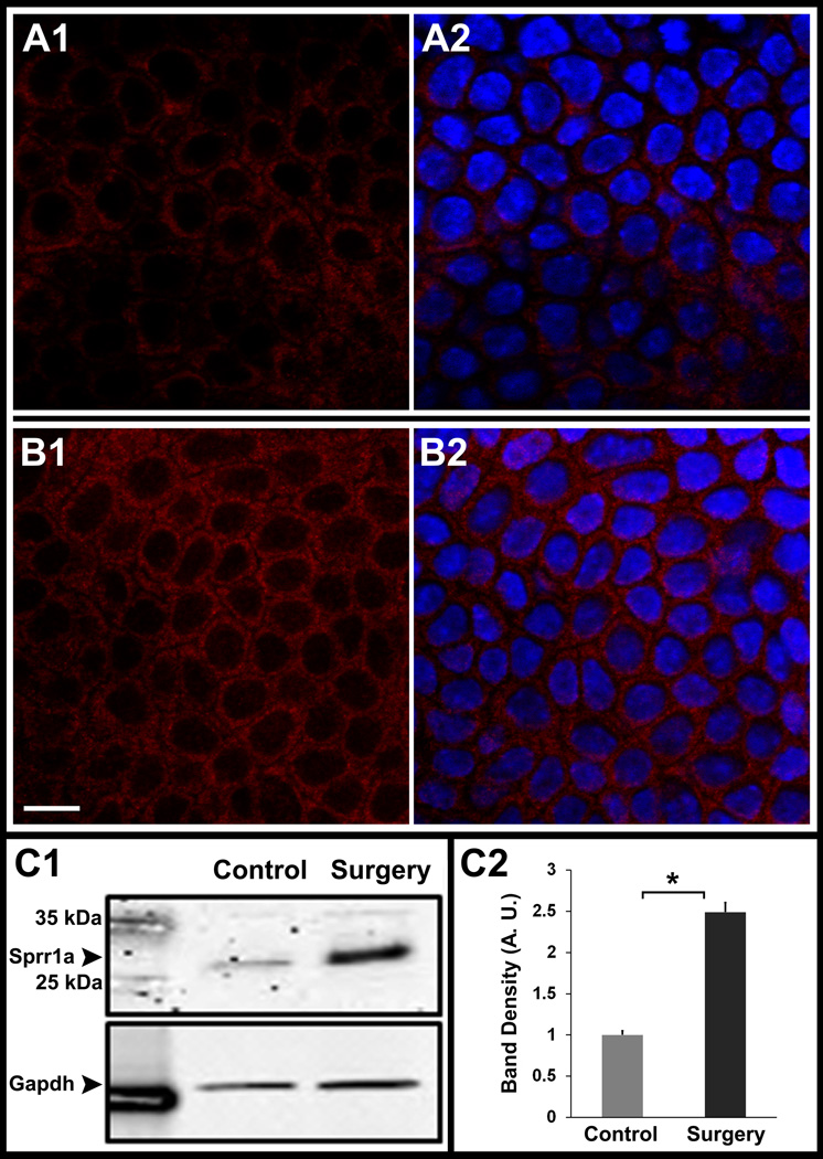
Confocal Immunofluorescent localization of Sprr1a in whole-mount corneas from thy1-YFP mice. (A,B) and Western analyses of corneal lysates for Sprr1a (C). Images show confocal sections in normal unoperated corneas (A1 – A2) and corneas at 2 weeks after lamellar transection (B1 – B2). In unoperated corneas, Sprr1a (red) is localized to corneal epithelial cell membranes (A1). Similarly, in operated corneas Sprr1a (red) is also localized to corneal epithelial cell membranes (B1). Image A2 and B2 shows an overlay with DAPI stained nuclei (blue). Using the same settings on the confocal microscope, the Sprr1a fluorescence in epithelium of operated corneas (B1) was greater than the epithelial fluorescence in unoperated control corneas (A1). Western analyses for Sprr1a (34 kDa) showed a denser band in operated corneas (C1, surgery lane) that was 2.48 fold higher than in unoperated corneas (C2). Gapdh (37 kDa) was used as a loading control. Scale bar, 10 µm. Asterisk, p < 0.05. Error bars, standard error of mean (SEM).