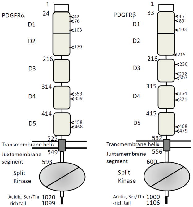
Fig. 4.
The two types of PDGFRs and their domain compositions. The starting numbers of the specific domains/segments in the coding sequences are marked left to the boundaries. Shown are all numbers for human PDGFRs. The positions of N-linked glycosylations are also marked. The lipid bilayer is represented by two straight lines. Note that D1 and D2 are an integral module, and the intracellular kinase domain is a split domains with an insert between N-terminal and C-terminal lobes.

