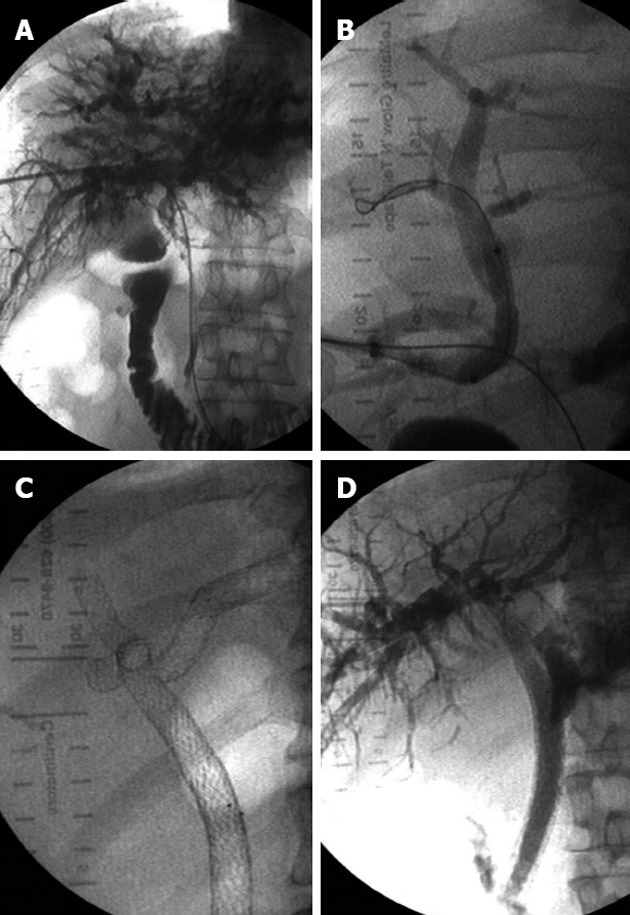
Figure 2.

Bismuth IV lesions. A: Percutaneous trans-hepatic cholangiography image demonstrating a Bismuth IV biliary obstruction; B: Right-sided approach; C: Multiple (n = 4) bilateral stenting in segmental ducts communicating with the stented common bile duct; D: Final fluoroscopic image demonstrating the flow of contrast medium into the duodenum.
