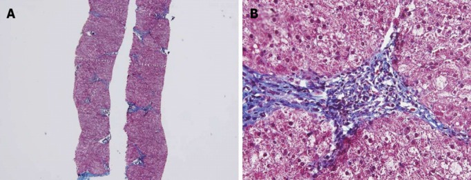
Figure 1.

Histological findings. Biopsy specimen showing mild lobular activity, mild portoperiportal activity and periportal fibrosis with frequent portal to portal bridging fibrosis. A: Masson’s Trichrome stain, 40×; B: Masson’s Trichrome stain, 400×.
