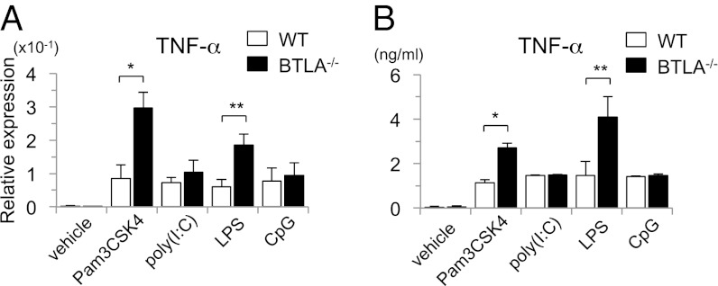
Fig. 3.
BTLA−/− DCs produce large amounts of TNF-α on stimulation with LPS and Pam3CSK4 but not poly(I:C) or CpG. BTLA−/− BMDCs and WT BMDCs were stimulated with LPS (100 ng/mL), Pam3CSK4 (200 ng/mL), poly(I:C) (10 μg/mL), and CpG (10 μg/mL). (A) Four hours later, the expression levels of TNF-α mRNA were measured by quantitative PCR. Data are mean ± SD, n = 4. *P < 0.05, **P < 0.01. (B) Twelve hours later, the levels of TNF-α in the supernatants were measured by ELISA. Data are mean ± SD, n = 4. *P < 0.05, **P < 0.01.

