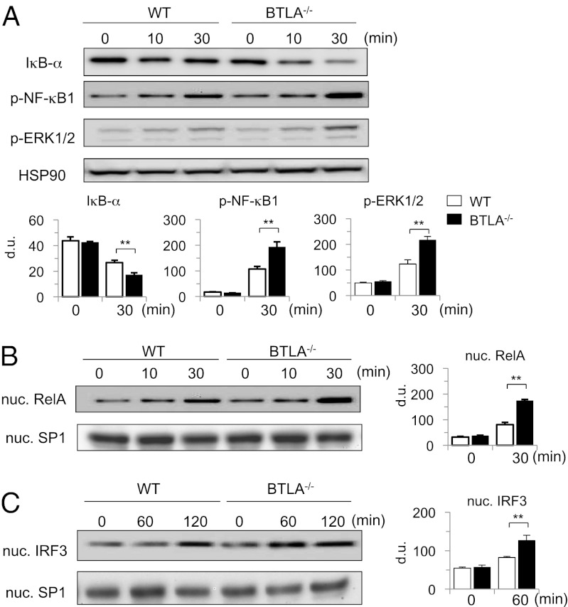
Fig. 4.
LPS-induced activation of NF-κB, MAPK, and IRF-3 pathways is enhanced in BTLA−/− DCs. (A and B) BTLA−/− BMDCs and WT BMDCs were stimulated with LPS (100 ng/mL) for indicated time periods. (A) Whole-cell lysates were subjected to immunoblotting with antibodies against IκB-α, p-NF-κB1, p-ERK1/2, and HSP90α/β (as a control). (B) Nuclear extracts were subjected to immunoblotting with antibodies against RelA and SP1 (as a control). Shown are representative blots and densitometric analyses of relative intensity of three independent experiments. (C) BTLA−/− BMDCs and WT BMDCs were stimulated with LPS (1 μg/mL) for indicated time periods. Nuclear extracts were subjected to immunoblotting with antibodies against IRF-3 and SP1. Shown are representative blots and densitometric analyses of relative intensity of three independent experiments. ** P < 0.01.

