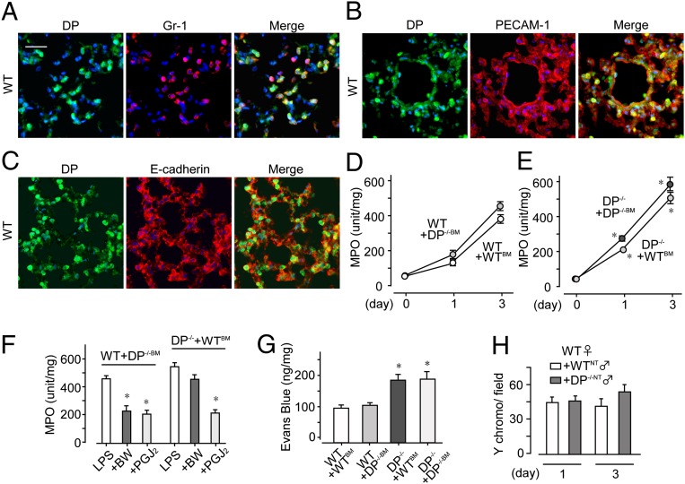
Fig. 5.
DP activation in endothelial cells is beneficial against ALI. (A–C) DP protein expression was detected in neutrophils (A) and endothelial cells (B), but not in epithelial cells (C), on day 3 (n = 5 each). (Scale bar: 50 μm.) (D and E) After bone marrow transplantation, MPO activity was monitored in WT, DP−/−, and CRTH2+ mice (n = 8 each). (F and G) BW245C or 15d-PGJ2 was administered to LPS-challenged mice, and MPO activity (F; n = 6–8) and dye extravasation (G; n = 6–8) were monitored on day 3. (H) The infiltrating ability of DP−/− neutrophils isolated from male mice into inflamed WT lung was monitored on day 3 (n = 8 each). *,†P < 0.05 compared with WT+WTBM (E), LPS-treated (F), and WT+WTNT (G) mice.

